2.3 From X-Rays to Computed Tomography
2.3.1 X-Rays and Computed Tomography: An Introduction
 |
|
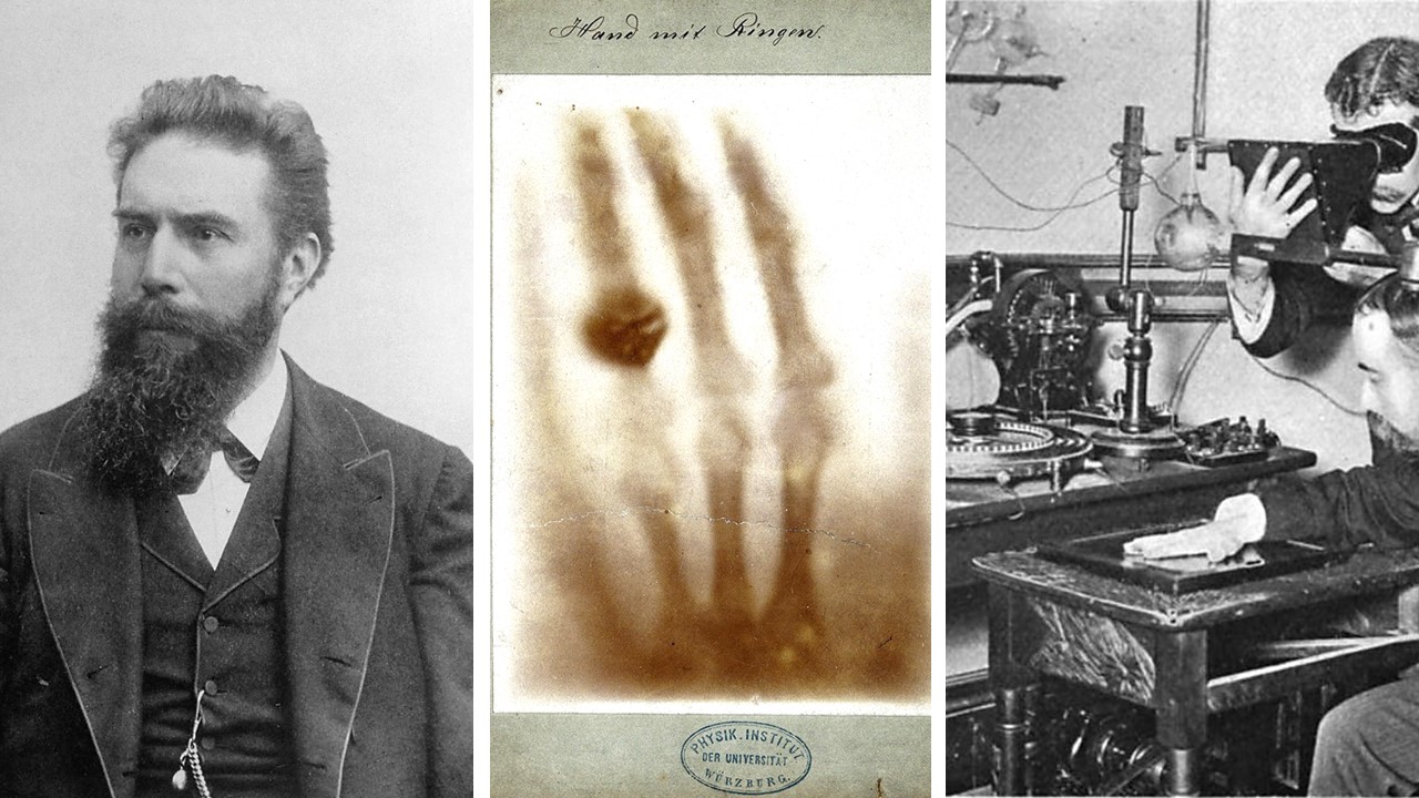
Wilhelm Conrad Röntgen and the First X-Ray; the hand of his wife, Anna Bertha (Image from the Röntgen Museum). On the right is an X-Ray taken in c. 1896. Click image to enlarge. |
 |
|
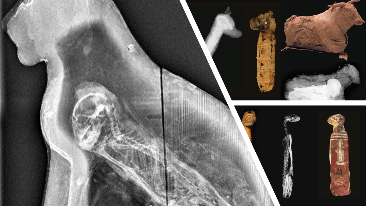
X-Rays of a cat sarcophagus (left) from the Manchester Museum, University of Manchester; of a mummified cat (bottom right) and of a dog and a bovine (top right) from The Brooklyn Museum Sources: Kahn, E. M. (2015). Animal Mummies Unwrapped. The New York Times. Oct. 2, 2015, Section C, Page 27 ttps://www.nytimes.com/2015/10/02/arts/design/animal-mummies-unwrapped.html / Eric A. Powell (2014). Messengers to the Gods. Archaeology Online, March/April 2014. https://www.archaeology.org/issues/124-1403/features/1724-egypt-animal-mummies-brooklyn-museum |
Computed Tomography was invented by Godfrey Hounsfield, who used to tinker as a child by taking apart electronics and devices. As he said in at the time of his Nobel Prize award:
At a very early age I became intrigued by all the mechanical and electrical gadgets which even then could be found on a farm; the threshing machines, the binders, the generators. But the period between my eleventh and eighteenth years remains the most vivid in my memory because this was the time of my first attempts at experimentation, which might never have been made had I lived in a city...I constructed electrical recording machines; I made hazardous investigations of the principles of flight, launching myself from the tops of haystacks with a home-made glider; I almost blew myself up during exciting experiments using water-filled tar barrels and acetylene to see how high they could be waterjet propelled (Nobel Lectures 1979).
As an electrical engineer, Hounsfield joined the EMI Laboratories after WWII. His invention of CT Scanning was based on the idea that the contents of a box can be determined by taking X-Rays around an object. Although his idea was originally rejected by many radiologists, Dr. James Ambrose, collaborated with him, also providing him with samples to test his prototype on. In the early 1970s, only head scanning prototypes were developed, while in 1976 body scanners were also produced and distributed to medical facilities in England and the USA. In his 1973 publication at the British Journal of Radiology, Hounsfield describes CT scanning:
a technique in which X-ray transmission readings are taken through the head at a multitude of angles: from these data, absorption values of the material contained within the head are calculated on a computer and presented as a series of pictures of slices of the cranium. The system is approximately 100 times more sensitive than conventional X-ray systems to such an extent that variations in soft tissues of nearly similar density can be displayed. (p. 1016)
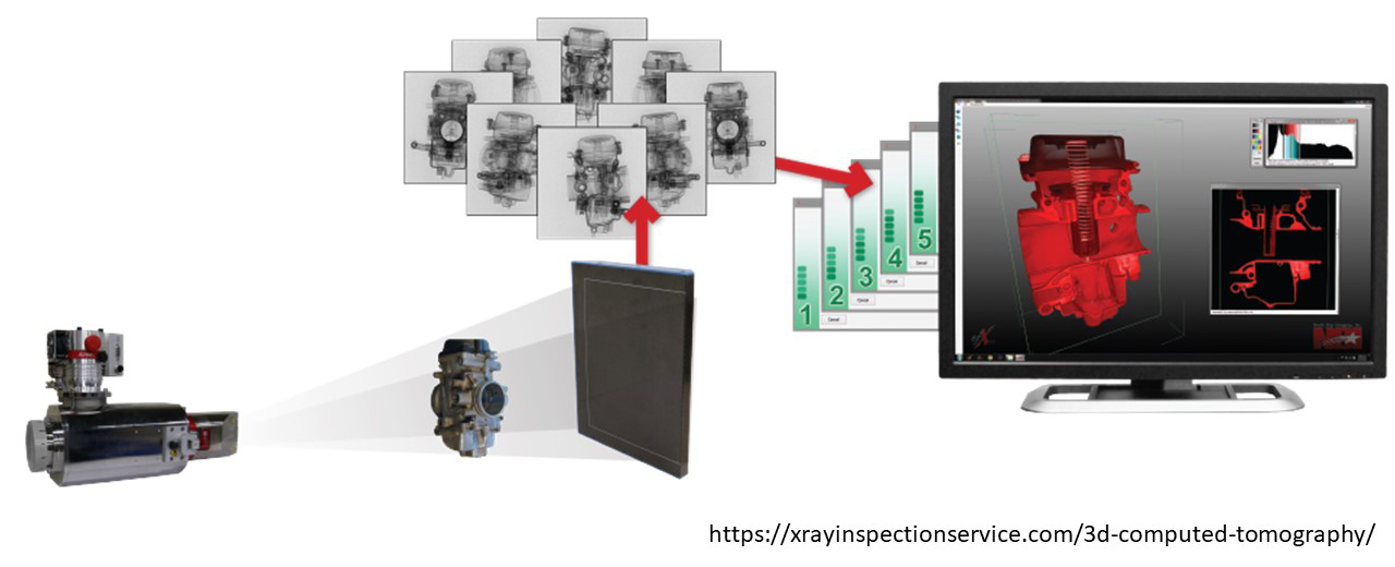
Contrary to X-Rays, which can only produce a two-dimensional representation of the scanned object/body, Computed Tomography (from the Greek tomos meaning 'slice' and grapho meaning 'describe') produces volumetric data, which can be used to generate 3D models. CT works similarly to X-Ray devices; there is an X-Ray source and X-Ray detectors opposite the X-Ray source. The device takes hundreds or even thousands of scans (2D digital radiography projections) in a 360-degree rotation around the object. In other words, the object is scanned in consecutive slices which together form a complete image of the object in cross-section. In a post-scanning phase, the 2D images are processed and reconstructed in a three-dimensional volume, therefore allowing the observation of the object at any 'slice' and angle (Applbaum & Applbaum 2005, p. 233). According to Hughes (2011, p. 60), CT scanning is ideally suited for heritage artefacts, since techniques such as Magnetic Resonance Imaging (MRI) require enough moisture to work, which in most cases is not preserved in such objects.
CT Scanning, although mostly used in medicine, as a diagnostic tool, has become very popular in cultural heritage (and beyond, e.g. in engineering) due to its non-destructive nature. It is a relatively and extremely accurate technique, which however, is not always easy to be applied to heritage artefacts. Such equipment, can typically only be found in hospitals and medical laboratories, as its very high cost of obtaining and maintaining it, make it unaffordable for non-medical related applications. There have been many cases, where heritage projects develop collaborations with medical facilities, but of course, the bureaucracy involved in transporting the artefacts (e.g. from a museum) to such a facility, often poses insurmountable difficulties. In the following page, you will explore some characteristic case studies for a range of materials and object types that demonstrate the potential of the method in the study of material culture.
References
- Applbaum, N., & Applbaum, Y. H. (2005). The use of medical computed tomography (CT) imaging in the study of ceramic and clay archaeological artifacts from the ancient near east. In Uda M., Demortier G., Nakai I. (eds). X-rays for Archaeology, pp. 231-245. Springer. https://doi.org/10.1007/1-4020-3581-0_17
- Hughes, S. (2011). CT scanning in archaeology. In Saba, L. (ed.) Computed tomography: Special Applications, pp. 57-70. InTech. https://www.researchgate.net/profile/Stephen_Hughes10/publication/221920121_CT_Scanning_in_Archaeology/links/55d5431208ae6788fa352c83.pdf
- Nobel Lectures (1967). Physics (1901-1921) - Wilhelm Conrad Röntgen – Biographical. Elsevier Publishing Company. https://www.nobelprize.org/prizes/physics/1901/rontgen/biographical
- Nobel Lectures (1979). Physiology or Medicine 1979 - Allan M. Cormack, Godfrey N. Hounsfield – Biographical. Elsevier Publishing Company. https://www.nobelprize.org/prizes/medicine/1979/hounsfield/biographical/
- The Nobel Prize in Physics 1901. NobelPrize.org. Nobel Media AB 2020. Sun. 17 May 2020. https://www.nobelprize.org/prizes/physics/1901/summary/
Further Reading
-
Beckmann, E. C. (2006). CT scanning the early days. The British Journal of Radiology, 79:5-8
https://doi.org/10.1259/bjr/29444122 - Hounsfield, G. N. (1973). Computerized transverse axial scanning (tomography): Part 1. Description of system. The British journal of radiology, 46(552), 1016-1022. http://qmxmt.com/scans/dave/other/papers/general%20tomography/hounsfield_1973.pdf
- Hughes, S. (2018). Computed Tomography. The Encyclopedia of Archaeological Sciences, 1-4. https://doi.org/10.1002/9781119188230.saseas0107
- O'Brien, J. J., Battista, J. J., Romagnoli, C., & Chhem, R. K. (2009). CT imaging of human mummies: a critical review of the literature (1979–2005). International Journal of Osteoarchaeology, 19(1), 90-98.
- Uda, M., Demortier, G., & Nakai, I. Eds. (2005). X-rays for Archaeology. Springer.


